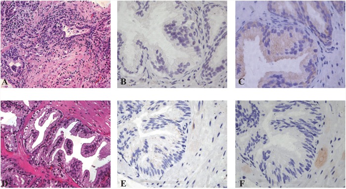
Figure 1. Staining of LC3B and P62 in prostate glands in different inflammatory conditions.

A-C. Inflammation score 8 prostatitis (A, 20x) showing LC3B negative staining (B, 40x) and P62 dot-like positive staining score 3 (C, 40x). D-F. Inflammation score 8 prostatitis (D, 20x) LC3B dot-like positive staining +2 score (E, 40x) and P62 negative staining (F, 40X) are shown.
