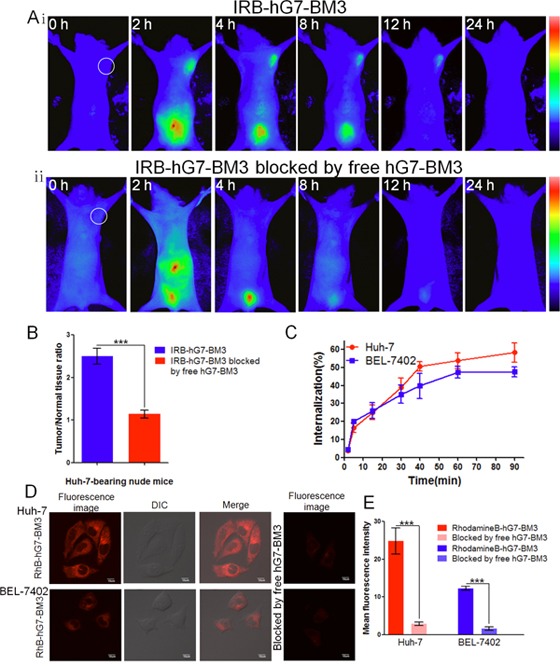
Figure 5. The tumor-targeting efficacy assay in vivo and tumor cell internalization assay in vitro.

(Ai) The bio-distribution of IRB-hG7-BM3 was evaluated by the NIR imaging assay in Huh-7-bearing nude mice within 24 h. (Aii) In blocking experiments, free hG7-BM3 inhibited the probes from binding to the tumor sites. (B) Tumor/normal tissue ratios calculated at 4-h post-injection of probe groups into Huh-7-bearing nude mice from the region of interest (ROIs). (C) hG7-BM3 internalized into hepatic cells rapidly within 40 min. The internalization rate was stabilizing at 90 min. (D) Laser confocal fluorescence microscopy images of Huh-7 and BEL-7402 cells incubated with the RhB-hG7-BM3 fluorescent probe, with or without a blocking dose of free hG7-BM3. (E) Mean fluorescence intensity of Huh-7 or BEL-7402 cells treated with RhB-hG7-BM3 probes, compared to blocking with free hG7-BM3. Data were given as the mean ± SD (n = 6). ***p < 0.001, ns: no significance.
