
Figure 2. Different fluorescence microscopy methods identify Sig1R inside the nucleus.
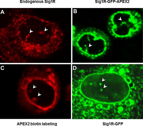
(A) Endogenous Sig1R was detected in NSC34 cells by immunocytochemistry using an in-house produced Sig1R antibody [40] with confirmed minimal non-specific labeling [7, 24]. (B) Sig1R-GFP-APEX2 fusion protein was expressed in a Sig1RKO NSC34 cell line (see Figure 1D). GFP fluorescence imaging indicates antibody-free Sig1R subcellular localization. (C) Sig1R-GFP-APEX2 fusion protein was expressed in Sig1RKO NSC34 cells followed by APEX2-catalyzed biotin labeling of proteins proximal to APEX2 (and hence Sig1R), whose localization was visualized via Cy3-labeled streptavidin. (D) Sig1R-GFP fusion protein was expressed in a Sig1RKO ARPE19 cell line (generated using the same method as in Figure 1D) and visualized by fluorescence microscopy. In all images, arrowheads indicate Sig1R localization inside the nucleus. Note the Sig1R-positive tubular nuclear structures within the nucleus.