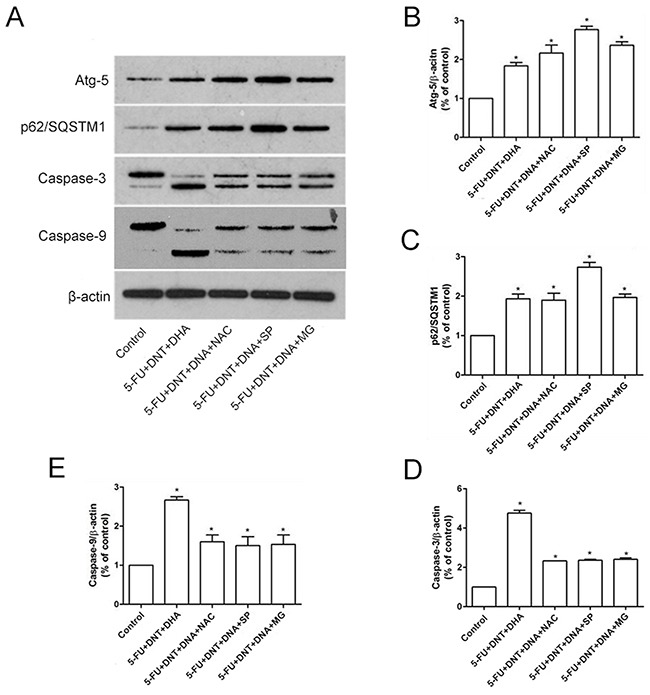
Figure 4. Western-Blot analysis of apoptosis- or autophagy- related proteins.

(A) The protein expression of Atg-5, p62/SQSTM1, Caspase-3 and Caspase-9 under different treatments were detected using western blotting. β-actin was used for loading control. (B-E) The densitometric analysis of Atg-5 (B), p62/SQSTM1 (C), Caspase-3 (D) and Caspase-9 (E) were exhibited in the right panels. *P <0.05 showed a significant difference compared to control.
