
Figure 9. Characteristics of Mcl-1, Caspase-3 and Caspase-9 expression in xenograft tumor mice.
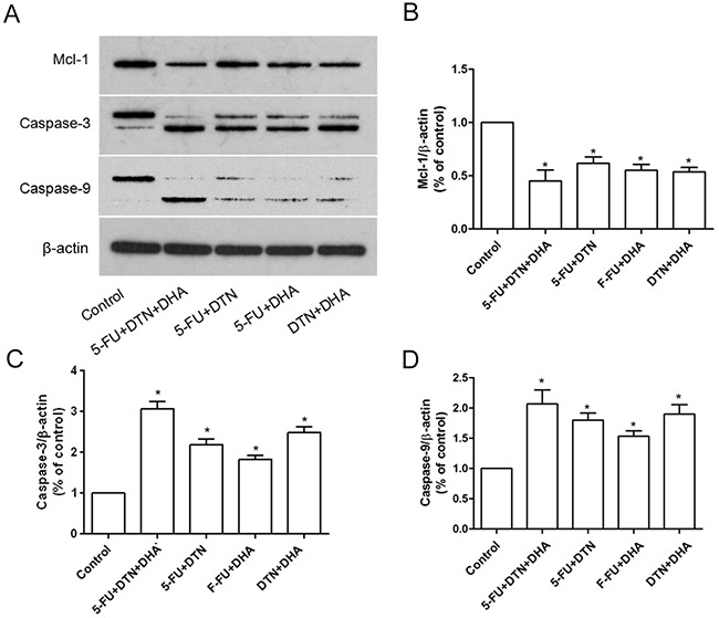
(A) The protein expression of Mcl-1, Caspase-3 and Caspase-9 in xenograft tumor from two or triple combination or 5-FU, DTN and DHA treated mice were detected using western blotting. β-actin was used for loading control. (B-D) The densitometric analysis of Mcl-1, Caspase-3 and Caspase-9 expression. *P <0.05 showed a significant difference compared to control.