
Figure 5. GT198+ pericytes in CD31+ vessel-derived oral tumor.
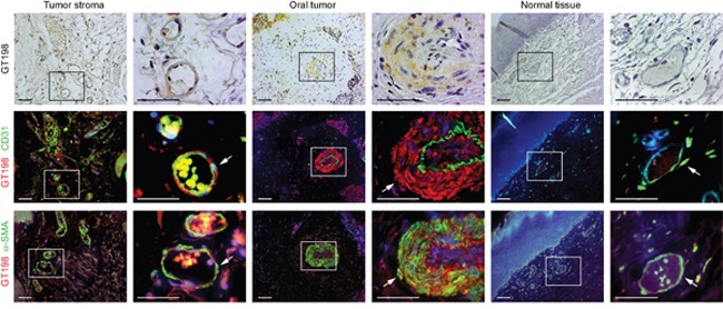
Three serial cut sections of oral tumor and adjacent normal tissues were analyzed for pericytes. Immunohistochemical staining of GT198 is shown in the top panels counter-stained with hematoxylin. Fluorescent double staining of GT198 in red, CD31 and α-SMA in green, and DAPI in blue, is shown in the middle and bottom panels, respectively. Each enlarged boxed area is shown at the right. In tumor stroma, GT198+ α-SMA+ pericytes enclose a thin layer of CD31+endothelial cells. In tumor, CD31+endothelial layer is enclosed by GT198+ pericytes, which further proliferate into tumor cells (Supplementary Figure 2). In normal vessels, GT198 expression is largely negative. Arrows indicate pericytes. Scale bars = 100 μm.