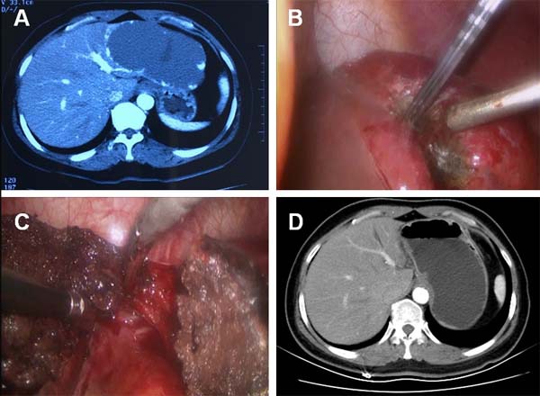
Figure 1. CT and intraoperative photos of case 1.

A. Contrast-enhanced CT demonstrated a 12.0-cm hepatic hemangioma located in the lateral left lobe of liver. B. Intraoperative photo showed the hemangioma at the resection margin shrank significantly after radiofrequency ablation. C. The hemangioma was removed and a 1.0-cm band of ablation-coagulated hemangioma was left in place. D. Contrast-enhanced CT showed that no residual tissue of the hemangioma exist in the tumor-dissected area.
