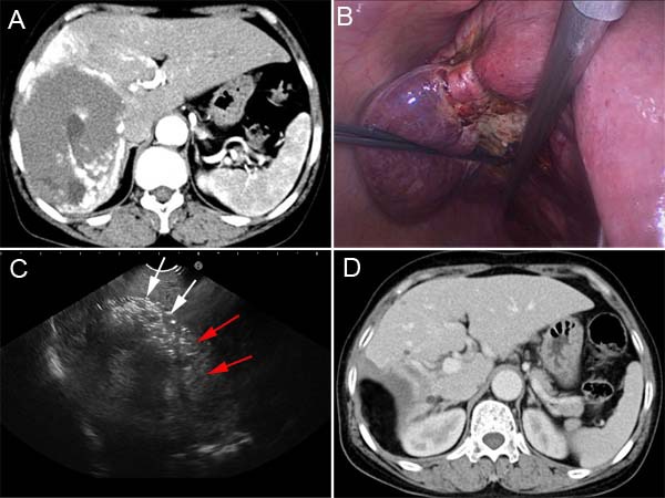
Figure 2. CT and intraoperative photos of case 2.

A. Contrast-enhanced CT demonstrated a 13.1 cm hepatic hemangioma in the right lobe. B. Radiofrequency ablation induced the significant shrinking of the hemangioma along the resection margin. C. Intraoperative ultrasound imaging was used to determine the boundary (red arrows) of hepatic hemangioma (white arrows) in liver parenchyma. D. Contrast-enhanced CT shows that the hemangioma was completely resected without residual tissue.
