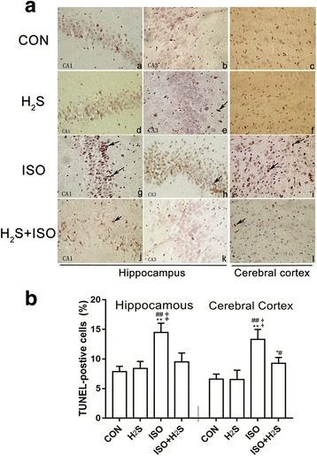
Fig. 1.

H2S attenuates isoflurane-induced neuro-apoptosis in hippocampus and cerebral cortex tissue. Paraffin sections were stained with the TUNEL technique using Apop Tag kit and counterstained with hematoxylin (A), under a high magnification (400×). For positive cells, nuclei stained dark brown with irregular or disintegration of different apoptotic bodies (indicated by the arrows); as for negative cells with nuclei light blue and regular. Quantification of apoptosis following TUNEL staining (B), calculating the apoptotic indices, expressed as Mean ± SD of five 400× fields in each rat (n = 6). * p < 0.05, ** p < 0.01 presented a significant difference compared with CON group; # p < 0.05, ## p < 0.01 presented a significant difference compared with H2S group, ‡ p < 0.01 presented a significant difference between ISO group and ISO + H2S group
