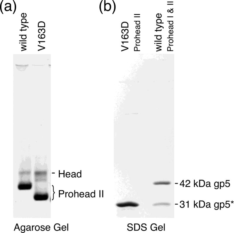
Figure 4. Gel analysis of wild-type and V163 mutant proheads.
(a) Agarose gel of Prohead II of wild-type and V163D mutant capsids. Each capsid protein was co-expressed with protease. After incubation, cells were lysed, and the lysate was processed through our prohead purification protocol. The resulting particles were analyzed on a 0.8% agarose gel. The heavy bands near the bottom of the gel are the proheads. The slower migrating lighter bands are spontaneously expanded proheads (i.e., heads). (b) SDS-polyacrylamide gel of the V163D mutant Proheads. The sample in the left lane is the V163D mutant Prohead preparation shown in lane 2 of the agarose gel in Figure 4. It displays a single subunit size. The right lane has a mixture of wild-type Prohead I and Prohead II, which are made of 42 kDa unprocessed gp5 or 31 kDa proteolytically processed gp5*, respectively. They serve as size standards and identify the subunits of the mutant prohead as the 31 kDa processed form of gp5.

