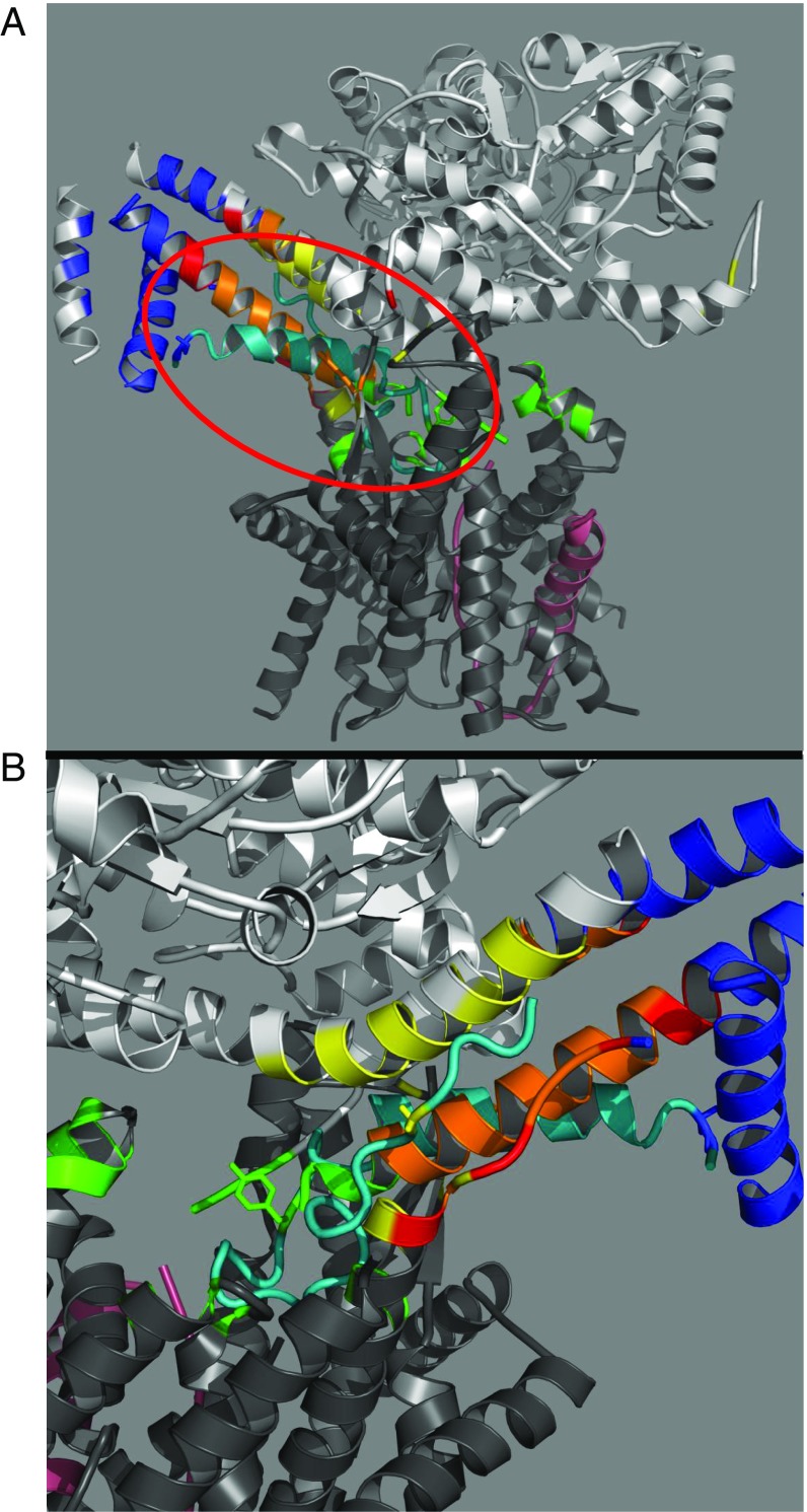
Fig. 3.
(A) FRET-mapped regions projected on the B. subtilis SecA–Geobacillus thermodenitrificans SecYE cocrystal structure (PDB ID code 5EUL). SecA is shown in light gray, SecYE is in dark gray, and the OmpA peptide substrate inserted at the end of the THF is shown in pink. For clarity, the nanobody crystallized with the complex has been omitted (10). Generation of FRET-mapped regions and their associated colors in the presence of ATP-γS was done as described in Fig. 2. Circled in red is the peptide substrate (residues 749–791) (shown in cyan) excised from the original 5EUL PDB structure and modeled into the mapped regions without any alteration of the original structure. (B) Enlarged view of the modeled peptide (cyan) and mapped locations. Residues 2 (Lys), 22 (Tyr), and 37 (Gly) of the OmpA peptide are shown in a stick representation in blue, green, and yellow, respectively, and exhibit excellent agreement with the PhoA-mapped locations. Note the adjacent C-terminal portion of SecY discussed in the text.

