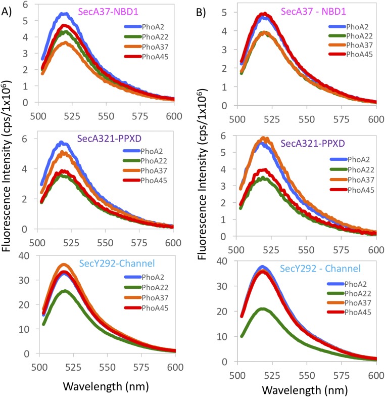
Fig. S2.
Fluorescence spectra of donor–acceptor labeled SecA–PhoA–SecYEG complexes generated with a 488-nm excitation wavelength. For all three sets of FRET pairs examined, the FRET efficiency is not linearly proportional to the position of the label on the PhoA portion of the chimera, consistent with a hairpin loop configuration. (A) All spectra were obtained in the presence of ADP. (Top) Spectra were generated with SecA37–AF647 as the acceptor, and the donor dye (AF488) was located at four different positions on the PhoA portion of the chimera: PhoA2 (blue), PhoA22 (green), PhoA37 (orange), or PhoA45 (red). (Middle) Spectra were generated with SecA321–AF647 as the acceptor and donor dye (AF488) positioned at either PhoA2 (blue), PhoA22 (green), PhoA37 (orange), or PhoA45 (red). (Bottom) The donor dye was located at SecY292–AF488, and the acceptor dye (AF647) was located at either PhoA2 (blue), PhoA22 (green), PhoA37 (orange), or PhoA45 (red). (B) Same as Fig. S2A, except all spectra were generated in the presence of ATP-γS. Spectral acquisition and analysis are described in SI Materials and Methods. FRET efficiencies, distances, and the degree of labeling are given in Tables S2–S4.

