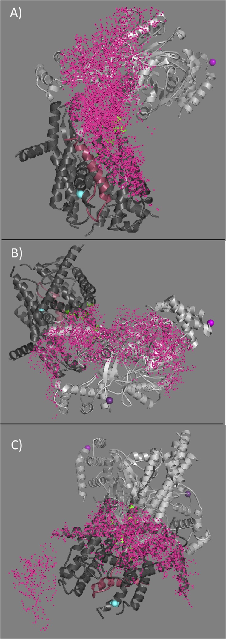
Fig. S4.
Depiction of the spherical shells (pink dots) used to identify the FRET overlap within the SecA–SecYE structure for PhoA residue 22 within the SecA–PhoA chimera. The shells were generated using distance values obtained in the presence of ATP-γS and are shown on the SecA–SecYE cocrystal structure PDB ID 5EUL. SecA is shown in light gray, SecYE is shown in dark gray, and the OmpA peptide is shown in pink. The width of the shells corresponds to the uncertainty in the FRET distance measurement (given in Tables S2–S4). (A) The shell determined from SecA residue 37 (magenta sphere) within NDB-1, (B) the shell determined from SecA residue 321 (violet sphere) within the PPXD, and (C) the shell determined from SecY residue 292 (cyan sphere) at the bottom of the channel. The intersection of the three spherical shells defines the region ascribed to PhoA residue 22 (green) in this case. The script for determining this intersection in given in SI Materials and Methods.

