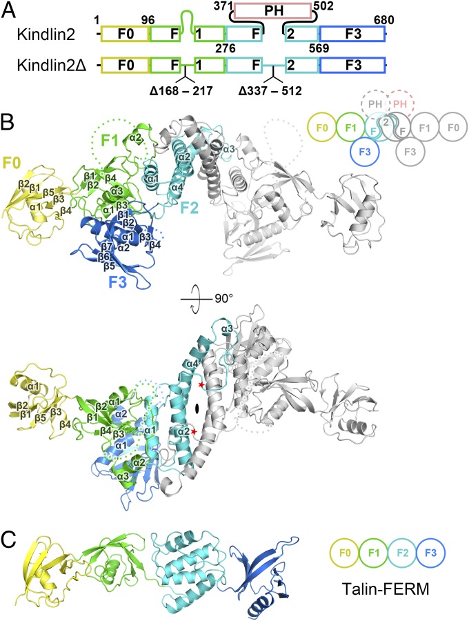
Fig. 1.
Overall structure of kindlin2. (A) The domain organizations of kindlin2. For structure determination, two regions were deleted as indicated in kindlin2Δ. The color coding of the regions is applied in all figures unless otherwise indicated. (B) Ribbon representation of the kindlin2Δ dimer structure. One protomer is colored follow the coding scheme in A, and the other identical protomer is colored in gray. The dimer is related by a twofold rotation axis, indicated by an ellipse. The disordered loops in the F1 and F3 lobes are indicated by hypothetical dotted lines. The PH domain deletion sites are indicated by red stars. The cartoon of the kindlin2 dimer schematically shows the interlobe interactions and the F2 domain-swapped dimer. (C) The talin-FERM structure (PDB ID code: 3IVF).

