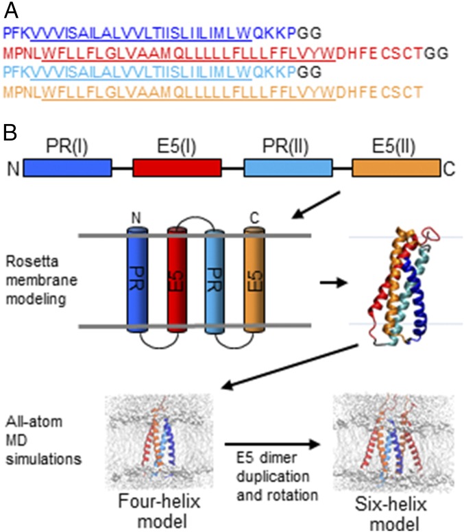
Fig. 1.
Modeling strategy. (A) Amino acid sequence of the virtual snake consisting of two TMDs of the PDGFβR and two TMDs of the BPV E5 protein. E5 sequences are orange [E5(I)] and red [E5(II)], PDGFβR TMD sequences are dark [PR(I)] and light [PR(II)] blue, and glycine linkers are black, with the predicted TMDs underlined. All sequences are written N-to-C. (B) Schematic overview of the multistep modeling strategy. Color scheme is as in A.

