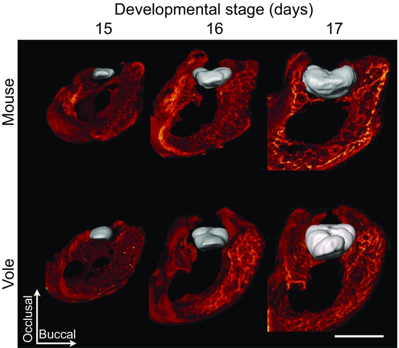
Fig. 3.
Soft tissue tomography reveals a close proximity of molars and jawbone from the onset of patterning. The first lower molar (m1) and its surrounding alveolar jawbone of mouse and vole are represented in false color (m1 in gray and jawbone in red) from the posterior view. An animation showing the development of the m1–jawbone relationship from E15 to E17 stages (Movie S1) further illustrates the close association of the jaw with growing tooth. Buccal is toward the right. (Scale bar, 500 µm.)

