Fig. 5.
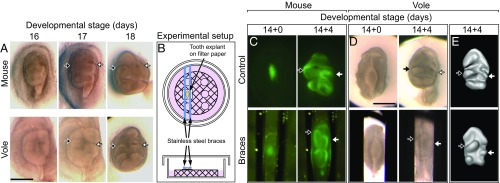
Cusp offset is lost in teeth cultured ex vivo and regained by using small braces. First lower molar (m1) explants of mouse and vole have largely parallel lateral cusp configurations (A). A Petri dish culturing system using small steel braces was used to constrain lateral expansion of teeth (B). ShhGFP-mouse molars show relatively normal cusp offset when cultured without braces and pronounced offset of the protoconid and the metaconid when cultured with closely spaced braces (C). Vole molars cultured with the braces rescue wild-type vole cusp offset. The cuspal patterns of vole molars (which lack GFP reporter activity) are more visible in 3D reconstructions made from soft tissue tomography (D and E). The protoconid is marked with a white arrow, and the metaconid with a black arrow. Anterior is toward the top. (Scale bar, 500 µm.)

