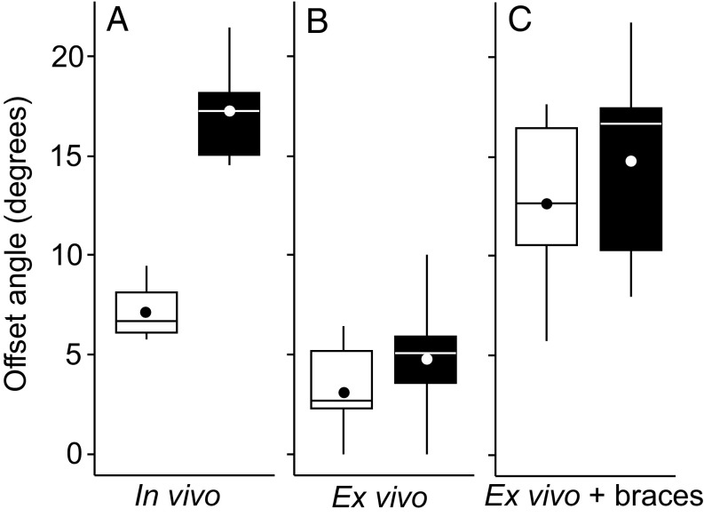
Fig. 6.
Both mouse and vole cusp offset can be controlled ex vivo. The mouse (white) and vole (black) cusp offset angles between the two first developing cusps, the protoconid and the metaconid, are distinct in vivo (n = 4 for mouse and 6 for vole) (A). In ex vivo cultured teeth, the cusps are almost parallel both in the mouse and the vole (n = 9 for mouse and 26 for vole) (B). In ex vivo cultured teeth with braces, the cusps show comparable offset both in the mouse and the vole (n = 6 for mouse and 5 for vole) (C). Boxes enclose 50% of observations; the median and mean are indicated with a horizontal bar and circle, respectively, and whiskers denote range. Statistical tests are in Table S5.

