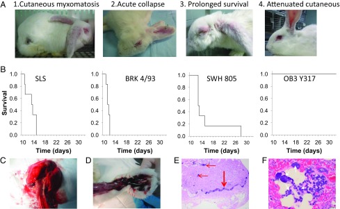
Fig. 1.
(A, 1–4) Typical clinical outcomes for clinical syndromes observed (Table 1 and SI Appendix). (1) Syndrome A (typical cutaneous myxomatosis). (2) Syndrome B (acute collapse with minimal signs of myxomatosis). (3) Syndrome C: progressive “amyxomatous” myxomatosis in a rabbit with prolonged survival. (4) Syndrome D (attenuated nodular cutaneous myxomatosis). (B) Kaplan-Meier plots for representative infections (SLS, 1950 progenitor virus, grade 1 virulence; BRK 4/93, 1993, grade 1 virulence; SWH 805, 1993, grade 2 virulence; OB3 Y317, 1994, grade 5 virulence). All rabbits recovered from the attenuated OB3 Y317 infection. (C) Pulmonary edema at autopsy. (D) Hemorrhage in hind leg at autopsy. (E) Histology of lymph node showing complete absence of lymphocytes and bacteria packed into subcapsular sinus (broad arrow) and other parts of the node (arrows). (Scale bar, 100 μm.) (F) Histology of lung showing edema and bacterial colonies (arrows). (Scale bar, 20 μm.)

