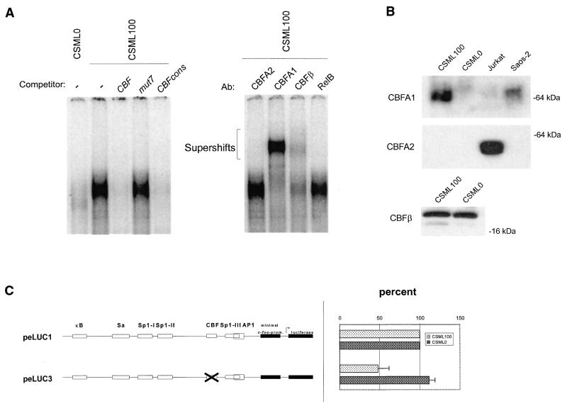
Figure 4.
Structural and functional analysis of the CBF-binding site. (A) EMSA of complexes formed by the CBF oligonucleotide with CSML0 and CSML100 nuclear extracts. As competitors the CBF oligonucleotide, mut7 or a CBF-containing oligonucleotide derived from the Moloney murine leukemia virus enhancer (CBFcons, Materials and Methods) were used. (Right) Supershift analysis of the CBF complex formed between CBF oligonucleotide and nuclear proteins from CSML100 cells. The competitors and antibodies used are indicated above the lanes. (B) Western blot analysis of proteins from CSML0 and CSML100 cells. The antibodies used for immunostaining are indicated on the left. Nuclear extracts prepared from Saos-2 and Jurkat cells expressing CBFA1 and CBFA2 proteins, respectively, were used as positive controls. (C) Transient transfection analysis of a reporter construct with a mutated CBF site in CSML0 and CSML100 cells.

