Fig. 2.
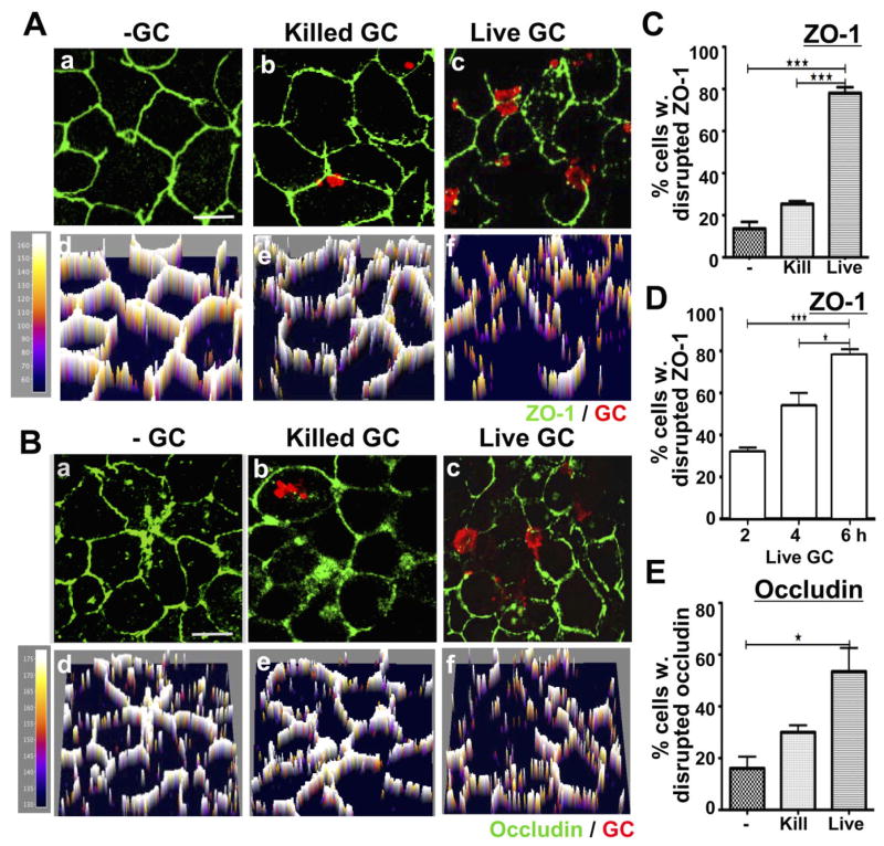
Viable but not killed GC disrupt the continuous apical junction location of ZO-1 and occludin in polarized HEC-1-B cells. Polarized HEC-1-B cells were incubated with media only (a and d), gentamicin killed GC (b and e) or live GC (c and f) in the apical compartment for 6 h (A–C and E) or for 2, 4 and 6 h (D). Cells were fixed and stained for ZO-1 (A, C and D) or occludin (B and E) and GC, and then analysed using confocal microscopy. Shown are representative images (composites of 1 μm slices) (a–c) and their fluorescence intensity profiles (d–f) at 6 h. The percentage of cells with disrupted ZO-1 (C) and occludin (E) peripheral staining at 6 h or with disrupted ZO-1 peripheral staining over time (D) was quantified by visual inspection, and the average percentages (± SD) from three independent experiments (~50 cells per experiment) are shown. Scale bar, 5 μm. ***P ≤ 0.001. *P ≤ 0.05.

