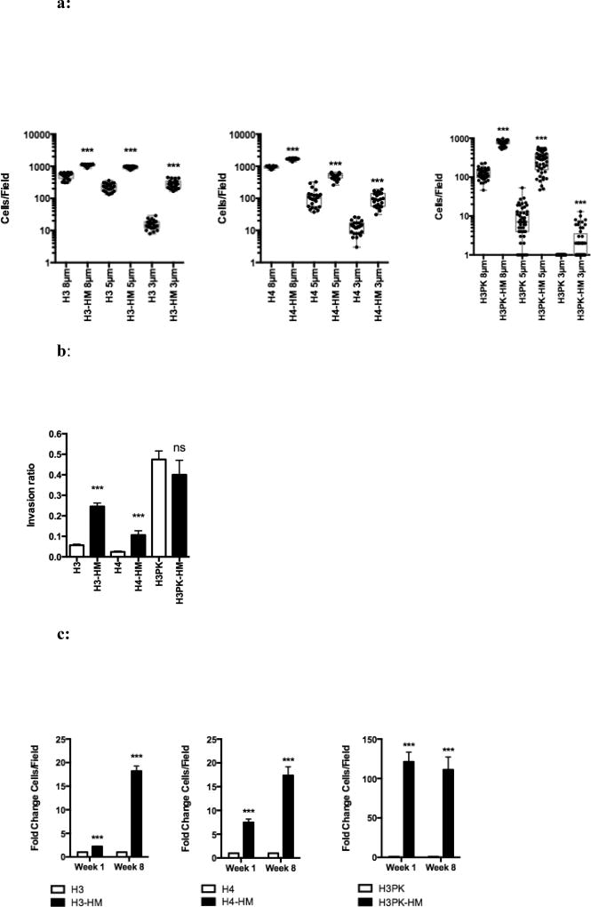
Figure 2.
a: Airway epithelial cells selected through increasingly smaller pores have enhanced migratory capacity. A transwell assay with basal growth medium in both chambers was used to determine cell migration. Migration quantified by fluorescence microscopy and ImageJ of at least 25 fields; each dot represents the number of cells observed in a single field. Mean ± SEM. ***, P < 0.001.
b: Highly migratory cells have characteristics that allow for enhanced invasion. The inverted invasion assay shows the invasion ratio (the number of cells invaded divided by number of non-invasive cells). Mean ± SEM. ***, P < 0.001.
c: The highly migratory phenotype remains stable eight weeks post-selection. The fold-change in the number of cells/field that migrated to the bottom chamber at week 1 (empty bars) and week 8 (filled bars) after selection and migration through 3 µm pores shown. At least 25 fields were analyzed. Mean ± SEM. ***, P < 0.001.

