Figure 5.
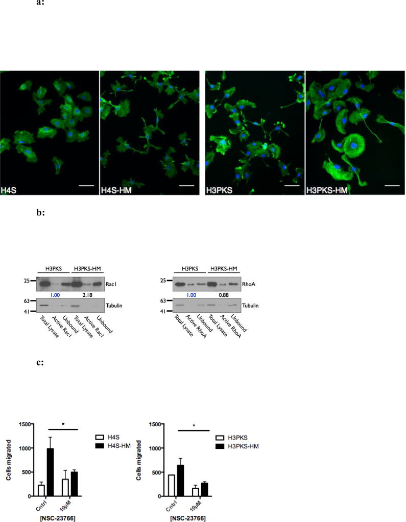
a: Actin filament structures are abundant in highly migratory cells that express SNAIL. Fluorescence micrographs showing phalloidin-stained F-actin pseudocolored green (nucleus counterstained with DAPI, blue). H3PKS-HM cells have ventral actin-arcs located in the lamellipodia, which are not observed in the unselected cells. H4S-HM cells show long cellular F-actin rich protrusions. 200× total magnification. Scale bar is 50 microns.
b: Highly migratory cells that over-express Snail have increased active Rac1. Western blot and corresponding densitometry showing the relative levels of activated (GTP-bound) RhoA and Rac1 GTPases.
c: High migratory rate facilitated in part through Rac1 activity. Treatment with NSC-23766, an inhibitor of Rac1 activation, perturbs migration through 5 micron pores in highly migratory cells. The absolute number of migrated cells was recorded in replicate wells. Mean ± SEM. *, P < 0.05.

