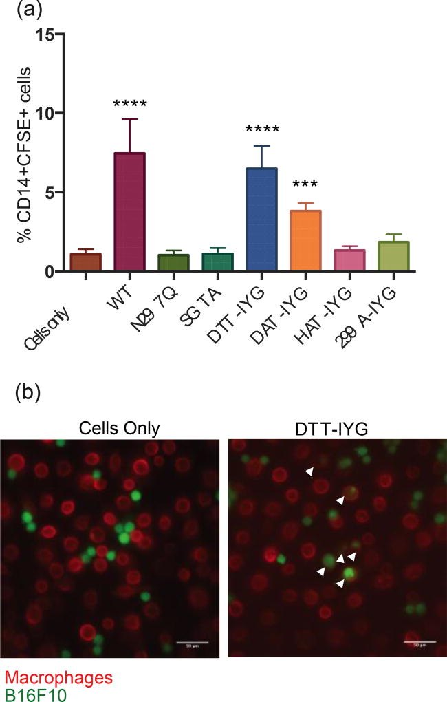
Figure 3.
Phagocytosis of tumor cells driven by aglycosylated antibodies. The negative control of “cells only” combine monocytes/macrophages with B16F10 tumor cells, which leads to a basal level of phagocytosis. Wild-type glycosylated antibody serves as the positive control. N297Q aglycosylated antibody serves as a second negative control. Macrophages are stained as CD14+ cells and B16F10 tumor cells as CFSE+. (a) Phagocytosis assay conducted from macrophages that were derived from monocytes harvested from whole human blood and matured with human GM-CSF. *** p-val < 0.001, **** p-val <0.0001 as compared to negative control of cells only (b) Microscopy images of phagocytosis of CFSE+ B16F10 cells (green) by CD14+ macrophages (red) at 20×. Arrows point to phagocytosis events. Scale bars at 50 µm.

