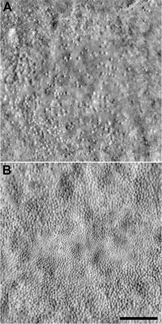
Figure 1.

Variability of the foveal cone mosaic in achromatopsia. Split-detector AOSLO images from the right eye of two different subjects with CNGB3-associated achromatopsia (and no cone function). (A) 16-year-old female with low peak cone density (9917 cones/mm2). (B) 37-year-old male with relatively high peak cone density (44,959 cones/mm2). Peak cone density was measured as reported by Langlo et al.17 Implications of this level of interindividual variability in remnant cone structure for defining the therapeutic potential of a given retina remain to be elucidated, though it is worth noting that the visual acuity of these two subjects was markedly different (20/800 for the subject in [A] and 20/100 for the subject in [B]). Scale bar: 100 μm.
