Figure 2.

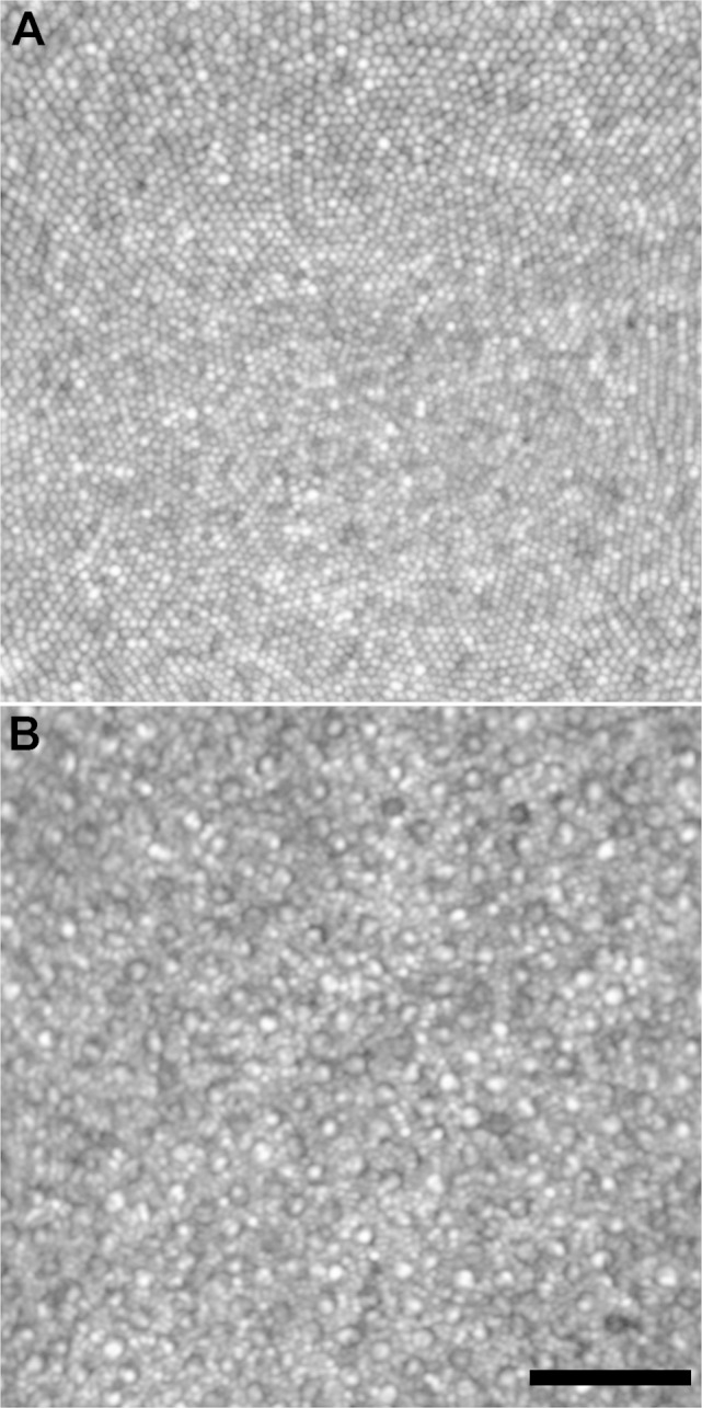
Confocal AOSLO images from a normal retina, displayed on a logarithmic scale. (A) Tightly packed cones in the fovea and (B) Cone and rod mosaic at 10° temporal to fixation. Right eye from a 27-year-old female (JC_11142). Scale bar: 50 μm.

Confocal AOSLO images from a normal retina, displayed on a logarithmic scale. (A) Tightly packed cones in the fovea and (B) Cone and rod mosaic at 10° temporal to fixation. Right eye from a 27-year-old female (JC_11142). Scale bar: 50 μm.