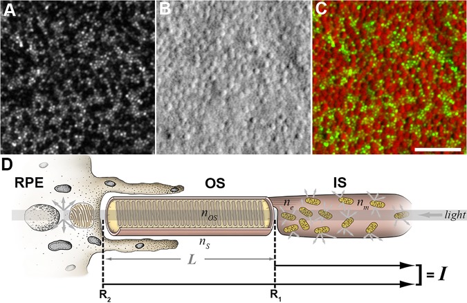
Figure 3.
Resolving cone inner and outer segment structure with AOSLO. Shown are confocal (A) and split-detection (B) images from the parafoveal retina of a patient with CNGA3-associated ACHM. The color-merged image (C) has the confocal image displayed in green and the split-detection image in red. Scale bar: 50 μm. (D) Photoreceptor schematic based off of a model presented by Jonnal et al.149 – the signal (I) requires intact photoreceptors42,149 and can vary as a result of small perturbations in photoreceptor structure. Multiply-scattered light from the RPE and inner segments is rejected by confocal AOSLO.

