
FIGURE 1.
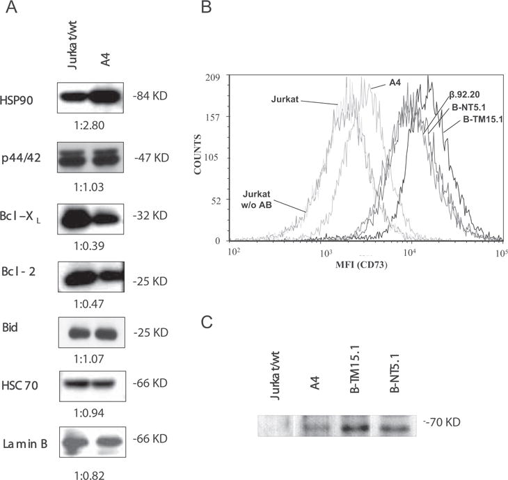
A, Immunochemical screening of antiapoptotic proteins. Lysates of parental Jurkat and A4 cells were subjected to Western blotting with Abs against HSP-90, ERK (p44/42), Bcl-2, Bcl-xL, and BID. Reactions were developed with HRP conjugates of corresponding secondary Abs and visualized by chemiluminescence. HSC70 and Lamin B were used as loading controls. Densitometry ratios are shown below the individual graphs. The densitometric value obtained with Jurkat cells equals 1. B, Analysis of CD73 expression by flow cytometry. The elevated CD73 expression in A4 cells was confirmed by FACS analysis. Resting Jurkat, A4 and different CD73 transfectant cells were labeled with anti-CD73 Ab 4G4, triple washed and visualized with secondary Abs conjugated to Alexa 647. The cells were washed once with PBS and data from 105 cells were collected on a FACScan (BD Biosciences) flow cytometer and analyzed with FCSExpress3 software. The following mean fluorescence intensity values were obtained from the FACS analyses: Jurkat wild-type labeled with nonspecific Ab, 2209; Jurkat wt, 2239; A4, 3672; β.92.20, 12294; B-NT5.1, 13345; B-TM15.1, 18289. C, Confirmation of CD73 expression on A4 cells by Western blotting. Lysates of 105 resting Jurkat and A4 cells equivalent to those shown in A were subjected to Western blotting with Abs against CD73. Lysates of 2 × 10⁴ Jurkat cells stably transfected with CD73 (B-NT5.1 and B-TM15.1) were used as positive controls.