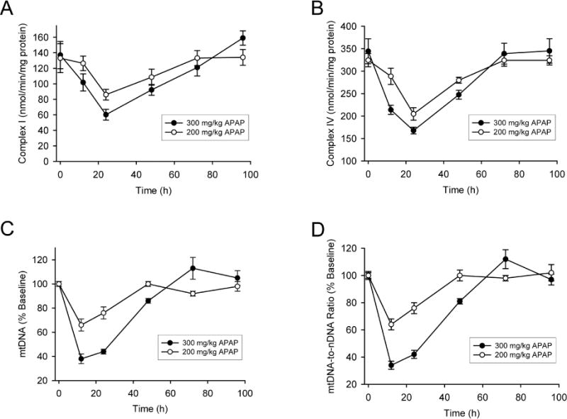
Figure 2. Electron transport chain activity and mitochondrial DNA levels in the liver after acetaminophen treatment.

Mice were treated with 200 or 300 mg/kg acetaminophen (APAP) and sacrificed at various time points between 0 and 96h. After isolation of liver mitochondrial by subcellular fractionation, enzyme activity of mitochondrial complex I (A) and complex IV (B) were then measured over time. Mitochondrial DNA (mtDNA) levels in the liver were also measured over time and expressed as absolute content (C) or normalized to nuclear DNA (D). Data are expressed as mean ± SEM for n = 4–6 animals per group and time point.
