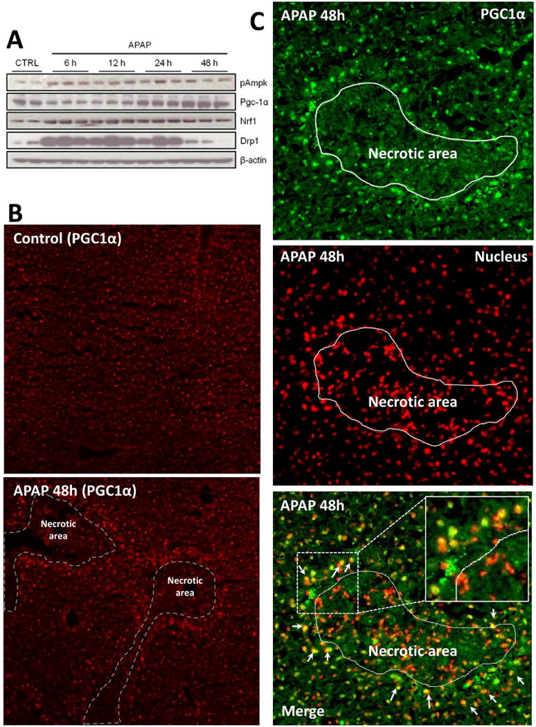
Figure 4. Activation or expression of mitochondrial biogenesis signaling mediators after acetaminophen treatment.

Mice were treated with 300 mg/kg acetaminophen (APAP) and sacrificed at various time points between 0 and 48h. (A) Liver homogenates were separated by SDS-PAGE and expression of several major mitochondrial biogenesis (MB) markers was assessed by immunoblotting. (B) Liver cryosections from controls and animals after 48h APAP were subjected to immunofluorescent staining of Pgc-1α (red). (C) For co-localization studies, in some liver sections 48h after APAP, Pgc-1α and nuclear DAPI signals were false colored green and red, respectively, to observe the merged signal as yellow (Original magnification 100×). Inset shows a magnification of the indicated area to show yellow merged signal in cell nuclei outside necrotic area. Necrotic areas were identified by pyknotic nuclei in DAPI images and marked as indicated.
