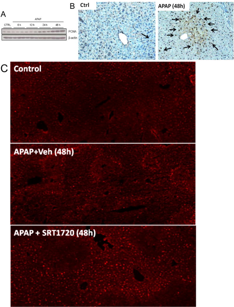
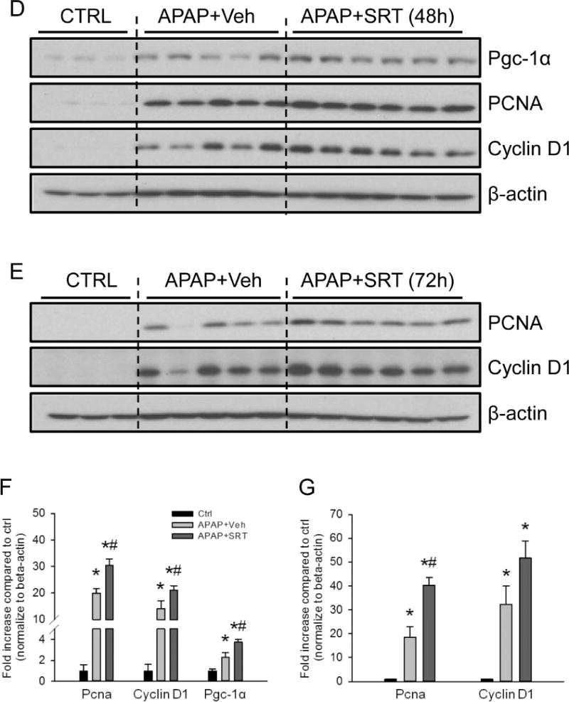
Figure 7. Induction of mitochondrial biogenesis promoted liver regeneration after acetaminophen treatment.


Mice were treated with 300 mg/kg acetaminophen (APAP). Some animals were treated with either SRT1720 (SRT) or its vehicle (Veh) as described in the materials and methods section, 12h and 36h post-APAP and sacrificed at 48h or 72h post-APAP. Following sacrifice, temporal expression of proliferating cell nuclear antigen (PCNA) was analyzed in liver homogenates by (A) western blotting, and (B) by immunohistochemistry on liver sections at 48h post-APAP (Original magnification 50×). Liver cryo-sections were also used for immunofluorescent staining of Pgc-1α in controls and animals treated with APAP ± SRT720 (Original magnification 100×). Pgc-1α (92 kD), PCNA (36 kD) and cyclin D1 (37 kD) expression were also examined by western blotting in livers from mice treated with SRT1720 (SRT) or its vehicle (Veh) at 48h (D, with densitometry in F) and 72h (E, with densitometry in G). Data are expressed as mean ± SEM for n = 3–5 animals per group and time point. *P< 0.05 vs. Ctrl. #P<0.05 vs. APAP + Veh.
