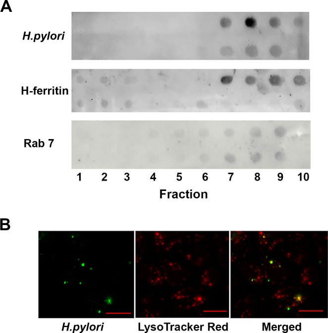
Fig 3. H. pylori co-localise with ferritin in Rab7-rich compartments within AGS cells.
(A) Phagosomes were collected from lysed AGS cells following infection with H. pylori strain 60190 (MOI, 10:1) for 15 h (top) or not (bottom) by sucrose density gradient centrifugation. Ten (top down) fractions were blotted onto PVDF membrane and probed with antibodies to H. pylori Lpp20, H-ferritin and Rab7. Antibody binding was visualised using a chemiluminescent detection of HRP-conjugated secondary antibody binding. Images are representative for three independent experiments. (B) AGS cells infected with DiO-labelled H. pylori strain 60190 and stained with LysoTracker Red are shown individually and in a merged image. Bar = 20μm.

