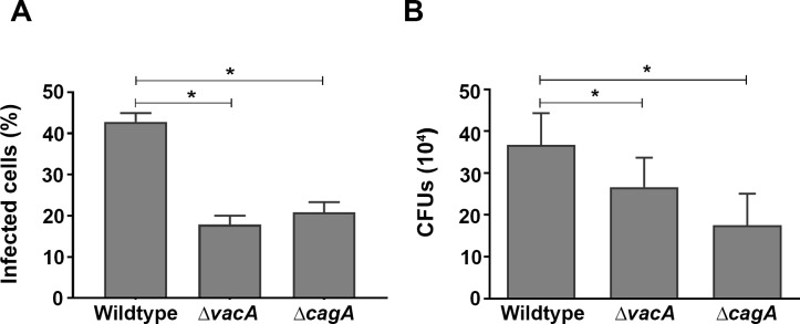
Fig 4. H. pylori uptake by AGS cells.
(A) Flow cytometry was used to assess internalization of DiO-labelled bacteria following infection of AGS cells with H. pylori (MOI, 10:1) for 15 h. (B) Infected cells were treated with gentamycin for 2 h to kill any remaining extracellular bacteria. Quantitative assessment of viable H. pylori in AGS cells was by serial dilutions of cells lysates. Results are ± SEM of three independent experiments. *, results are statistically different from those of cells infected with H. pylori wildtype strain 60190 (P<0.05), as determined by one-way ANOVA with Tukey’s test for multiple comparisons.

