
Figure 3.
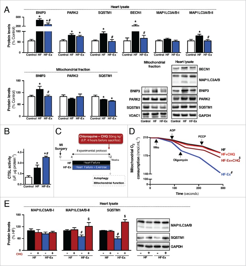
Exercise re-establishes autophagic flux in failing hearts. (A) Protein levels of BNIP3, PARK2, SQSTM1, BECN1 and MAP1LC3A/B in heart lysate and BNIP3, PARK2 and SQSTM1 in isolated mitochondria from sedentary sham-treated (control), sedentary heart failure (HF) and exercised heart failure (HF-Ex) rat hearts. (B) Cardiac CTSL activity in control, HF and HF-Ex rats. Data are presented as mean ± SEM.*, p < 0.05 vs. control; #, p < 0.05 vs. HF rats. (C) Schematic panel illustrating the study design: male Wistar rats were submitted to myocardial infarction (MI) surgery. 4 wk later rats were randomly assigned into sedentary heart failure (HF) and exercised heart failure (HF-Ex) groups. Exercised heart failure rats were trained on a treadmill over 8 wk. At the end of the protocol, 12 wk after surgery, rats were treated with a single intraperitoneal injection containing CHQ (50 mg.kg−1) to inhibit autophagic flux. Four h after injection animals were killed and autophagy-related markers and mitochondrial function were assessed. (D) O2 consumption in cardiac-isolated mitochondria and (E) protein levels of autophagy-related markers (MAP1LC3A/B and SQSTM1) in heart lysate from HF and HF-Ex rats treated with saline (−) or CHQ (+). Data are presented as mean ± SEM. #, p < 0.05 vs. HF (−) rats; $, p < 0.05 vs. HF-Ex (−) rats.