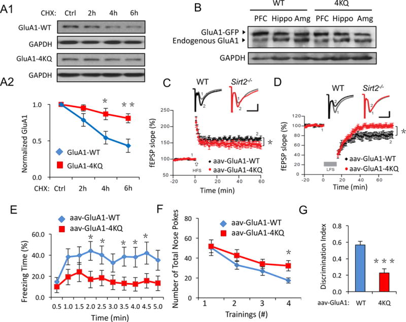
Figure 7. Mice with overexpression of acetylation mimetic AMPARs show defects in synaptic plasticity and memory.

(A) GluA1-WT, 4KQ degradation assays in transfected HEK cells. Compared to GluA1-WT, the GluA1-4KQ showed a reduced degradation rate (n = 3). (B) Adenovirus of GFP-tagged GluA1-WT or GluA1-4KQ was injected into the lateral ventricles of wild-type mice at P2 and brains were collected at P60. Western blot revealed high expression levels of viral GFP-GluA1 in brain regions including prefrontal cortex (PFC), hippocampus (Hippo) and amygdala (Amg). (C, D) Hippocampal brain slice recordings of LTP and LTD. GluA1-4KQ viral infected mice showed impaired LTP and LTD compared to the GluA1-WT control (n = 12 WT or 15 4KQ, p < 0.05). (E–G) Similar to the Sirt2−/− mice, GluA1-4KQ viral infected mice showed impaired contextual fear memory (E) and learning memory (F, G). AAV, adeno-associated virus. Bar graphs represent mean ± S.E., *P < 0.05, **P < 0.01, ***P < 0.001. Student’s two-tailed t test (A, E–G) and one-way ANOVA (C, D).
