Figure 1.
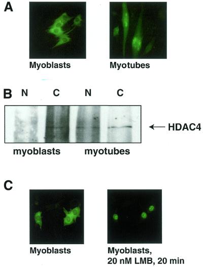
HDAC4 relocalises from the cytoplasm to the nucleus after myoblast fusion. (A) C2C12 myoblasts were microinjected with pcHDAC4-GFP. Four hours after injection living cells were visualised using fluorescence microscopy. To visualise HDAC4–GFP in myotubes, myoblasts were transferred to differentiation medium, incubated for 2 days and microinjected with pcHDAC4-GFP. Living myotubes were visualised using fluorescence microscopy. (B) Cytoplasmic and nuclear extracts were prepared from one 15 cm dish each of either 40–50% confluent myoblasts or myotubes after 5 days differentiation. Extracts were mixed with HDAC4-specific antibody (HD4B) and immunoprecipitated using protein A/G beads. Immunoprecipitates were separated using SDS–PAGE and western blotting was performed using a second HDAC4-specific antibody (HD4A). (C) C2C12 myoblasts were microinjected with pcHDAC4-GFP and living cells were visualised using fluorescence microscopy. Leptomycin B (LMB) was added to the medium at 20 nM and HDAC4–GFP fluorescence was monitored for 20 min.

