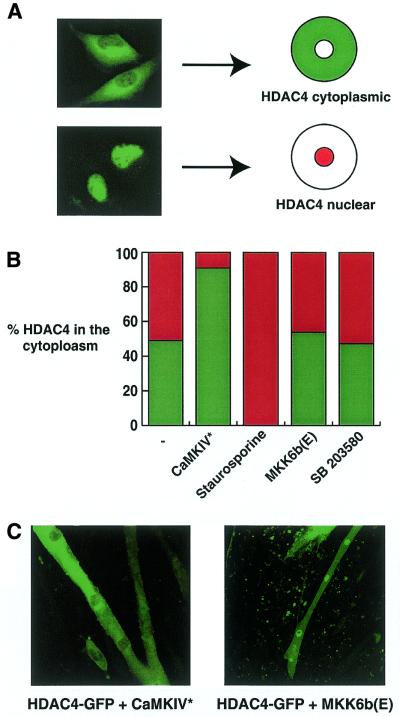
Figure 5.
Active CaMKIV, but not MKK6b, regulates HDAC4 localisation. (A) C2C12 myoblasts were transfected with 100 ng pcHDAC4-GFP. After transfection cells were transferred to differentiation medium for 24 h before fixing. GFP staining was visualised using confocal microscopy. HDAC4-GFP localised either to the cytoplasm or the nucleus. (B) C2C12 myoblasts were transfected with 100 ng pcHDAC4-GFP either in the presence or absence of pRSV-CaMKIV* or pMKK6b(E) (500 ng each). After transfection cells were transferred to differentiation medium for 24 h before fixing. Prior to fixing staurosporine (20 µM) or SB203580 (10 µM) was added to the culture medium for 1 h, as indicated. GFP staining was visualised using confocal microscopy. Percentages of cells displaying cytoplasmic staining are given (n > 200). (C) C2C12 myoblasts were transfected with 100 ng pcHDAC4-GFP either in the presence or absence of pRSV-CaMKIV* or pMKK6b(E) (200 ng each). After transfection cells were transferred to differentiation medium for 4 days before fixing. GFP staining was visualised using confocal microscopy.

