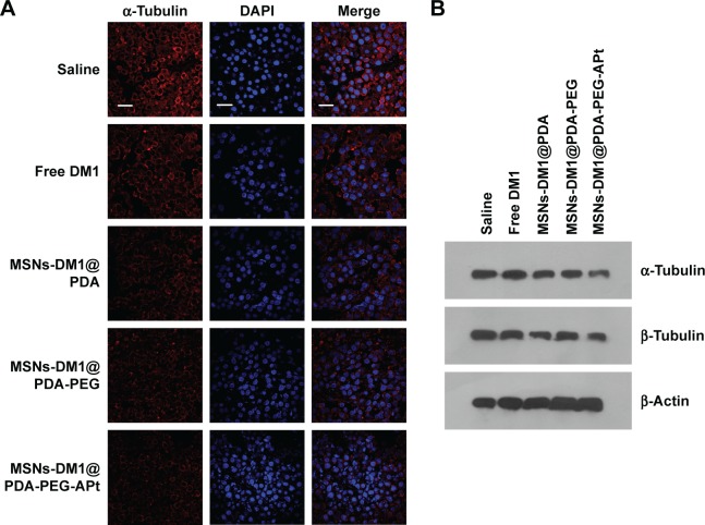
Figure 10.
Tubulin and apoptosis related proteins in tumor tissues from nude mice after different treatments. (A) Immunohistological staining with anti-α-tubulin antibody of tumor tissues. Microtubules (red) and nuclei (blue; DAPI) are shown, scale bar =10 µm. (B) Western blot analysis of α-tubulin in tumor tissues.
Abbreviations: MSNs, mesoporous silica nanoparticles; PDA, hydrochloride dopamine; PEG, polyethylene glycol; APt, aptamer; DAPI, 4′-6′-diamino-2-phenylindole hydrochloride.

