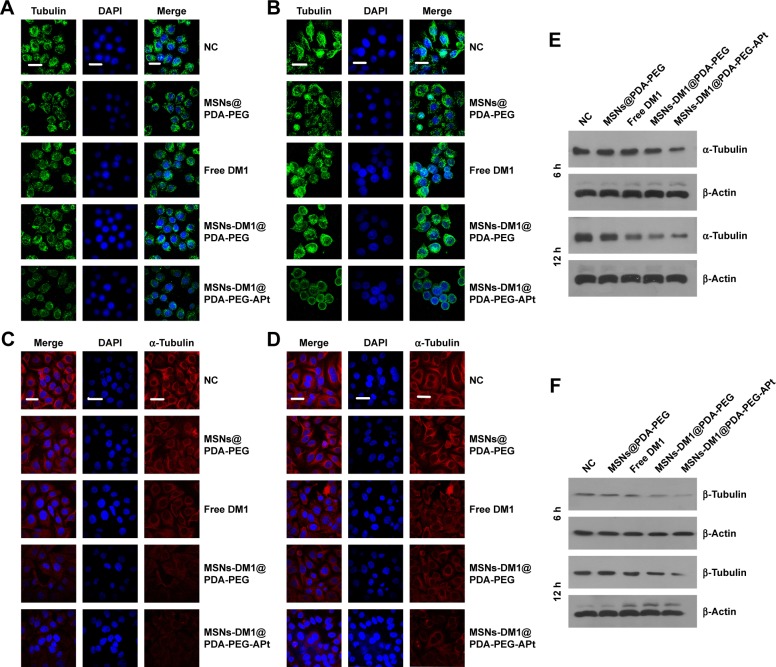
Figure 6.
SW480 cells treated with MSNs@PDA-PEG, free DM1, MSNs-DM1@PDA-PEG, and MSNs-DM1@PDA-PEG-APt for 6 and 12 hours, respectively. (A, B) Microtubule morphology was assessed with a microtubule fluorescence staining kit. Microtubules are stained green, and nuclei stained with DAPI (blue), scale bar =10 µm. (C, D) Cells were immunostained using anti-α-tubulin antibody to assess the effects of different DM1 formulations on α-tubulin. Red, α-tubulin; blue, nuclei (DAPI), scale bar =10 µm. (E, F) Western blotting was performed with antibodies against α-tubulin, β-tubulin, and β-actin at the indicated time points.
Abbreviations: NC, negative control; MSNs, mesoporous silica nanoparticles; PDA, hydrochloride dopamine; PEG, polyethylene glycol; APt, aptamer; DAPI, 4′-6′-diamino-2-phenylindole hydrochloride.

