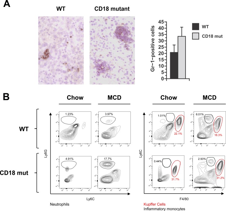
Fig 3. Hepatic inflammation in WT and CD18-mutant mice in response to MCD feeding.
(A) Gr-1 staining and quantitation of Gr1-positive cells in WT and CD18 mut liver at 3 wk. Photomicrographs illustrate that Gr-1-positive cells are abundant in both WT and CD18-mutant (CD18 mut) mice after MCD feeding, although in different distributions. Original magnification 15X. Histograms illustrated Gr1-positive cell counts in WT and CD18 mut livers, performed as described in Methods. Values represent mean ± SE for n = 5. (B) Representative FACS plots of hepatic leukocytes from mice fed chow or MCD diets. MCD feeding enhances the proportion of neutrophils (Ly6Ghigh) and inflammatory monocytes (Ly6Chigh), while the lack of CD18 further enhances the accumulation of neutrophils but not inflammatory monocytes. Data are illustrative of n = 6 mice per group (24 per cohort), performed as 2 replicate experiments involving 12 mice each (3 per group).

