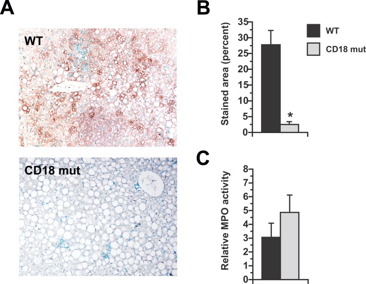
Fig 5. Chlorotyrosine-protein adduct formation in the livers of MCD-fed mice.
(A) Immunohistochemical staining for chlorotyrosine-protein adducts in WT and CD18 mut mice fed MCD diets for 3 wk. Original magnification 15X. (B) Morphometric quantitation of adduct-stained area. (C) Liver myeloperoxidase (MPO) activity normalized to the level in chow-fed WT mice. Values represent mean ± SE for n = 5. *P < 0.05 vs. WT.

