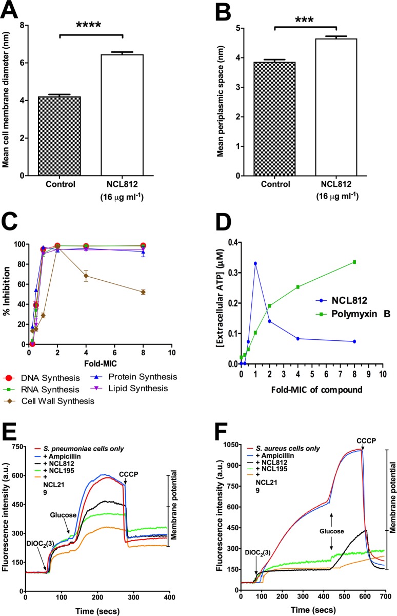
Fig 3. NCL812 compounds exert their antibacterial action on the cell membrane of S. pneumoniae and S. aureus.
(A and B), S. pneumoniae strain D39 exposed to 16 μg/ml NCL812 for 6 h exhibited significantly thicker cell membranes compared to untreated samples (A) (p < 0.0001; two-tailed unpaired t-test) and displayed significantly wider periplasmic space compared to untreated samples (B) (p < 0.001; two-tailed unpaired t-test. Data presented are an example from 12 different bacterial cells, each with at least 10 measurements per bacteria for both treated and untreated samples. (C and D) NCL812 affects macromolecular synthesis (c) and ATP release (D) in S. aureus. (E and F), NCL Compounds dissipate the membrane potential of S. pneumoniae and S. aureus. Membrane potential measurements of S. pneumoniae D39 (E) and S. aureus ATCC49775 (F). Bacterial suspensions were exposed to 16 μg/ml NCL812, NCL195, NCL219, or ampicillin (control) for 5 min after which DiOC2(3) was added and the fluorescence monitored until it plateaued. Cells were then re-energized with 0.5% glucose and the establishment of a membrane potential was measured as an increase in fluorescence until it plateaued. The membrane potential was then disrupted by the addition of the proton ionophore (CCCP). Data presented is representative of two experiments. For full description, see Materials and Methods.

