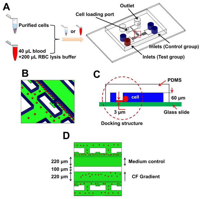
Fig. 3. Illustration of the microfluidic device for cell migration test.
(A) Microfluidic device illustration to test purified cells or directly from whole blood with RBC lysis. (B–C) Enlarged view of the microfluidic channel with cell docking. The red spheres indicate the cells, which were initially aligned by the thin barrier channel because the cell size is larger than the height of the barrier channel. Upon chemoattractant stimulation, cells will change their morphology to crawl through the barrier channel into the gradient channel. (D) Illustration of a cell migration experiment for parallel chemotaxis test and medium control. CF indicates chemotactic factor. Blue color indicates the channel walls and green color reflects the glass surface.

