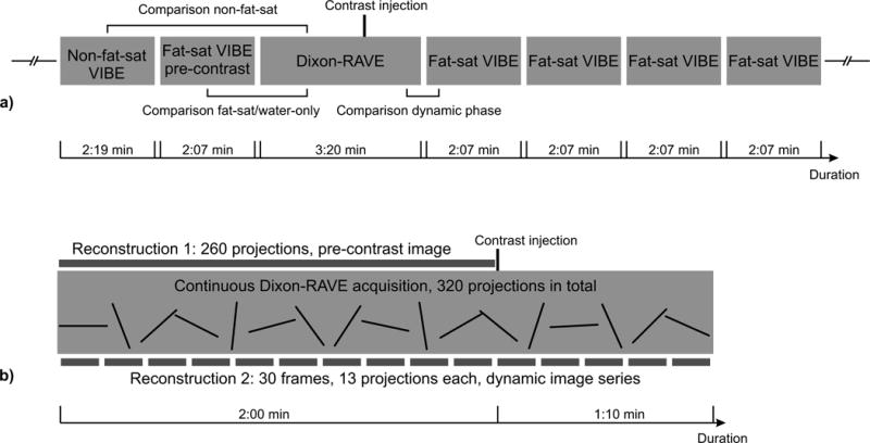
Figure 1.

a) Schematic illustration of the performed protocol, showing the different scans that were performed for each patient. Brackets indicate the comparisons that were used for the reader evaluation.
b) Overview of the two different Dixon-RAVE reconstructions. Reconstruction 1: To obtain static pre-contrast fat and water images, the first 260 projections were used. Reconstruction 2: For the dynamic image series, 13 consecutive projections were combined for each frame.
