Figure 3.

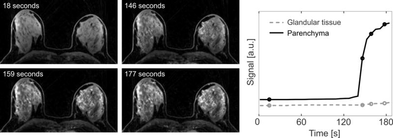
Dynamic water series and extracted enhancement curves, generated from Dixon-RAVE. The temporal resolution of 6.1 s allows clearly showing the marked background parenchymal enhancement.

Dynamic water series and extracted enhancement curves, generated from Dixon-RAVE. The temporal resolution of 6.1 s allows clearly showing the marked background parenchymal enhancement.