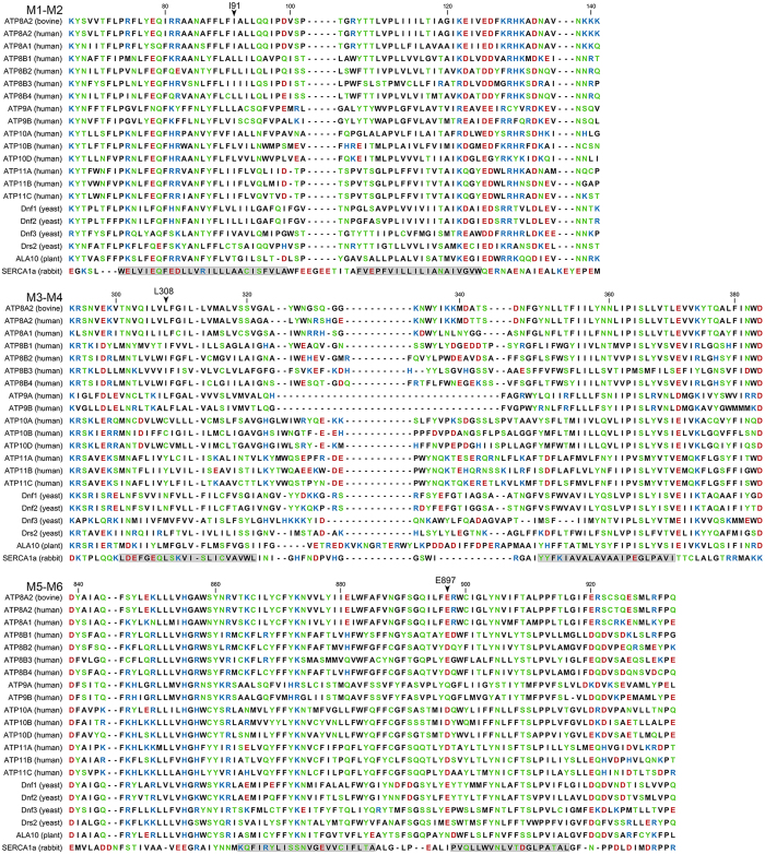
Figure 6.
Alignment of the regions corresponding to the first six transmembrane segments of P4-ATPases and SERCA. Protein sequences of the bovine ATP8A2 studied here, the 14 human flippases, 4 yeast flippases, and one plant flippase (UniProt nos. from above: C7EXK4, Q9NTI2, Q9Y2Q0, O43520, P98198, O60423, Q8TF62, O75110, O43861, O60312, O94823, Q9P241, P98196, Q9Y2G3, Q8NB49, P32660, Q12675, Q12674, P39524, and Q9LI83) were aligned with rabbit SERCA1 Ca2+-ATPase (UniProt no. P04191) as previously described14. Letter colours denote positively (blue) or negatively (red) charged, polar (green), or hydrophobic (black) residues. The residues replaced in the three disease mutants studied here are indicated with arrowheads. Numbering refers to the bovine ATP8A2. Grey shading denotes the transmembrane helices identified in SERCA crystal structures. The human P4-ATPases are subdivided into classes of genetically most related proteins: Class 1a (ATP8A1, ATP8A2), Class 1b (ATP8B1, ATP8B2, ATP8B3, ATP8B4), Class 2 (ATP9A, ATP9B), Class 5 (ATP10A, ATP10B, ATP10D), and Class 6 (ATP11A, ATP11B, ATP11C)2.

