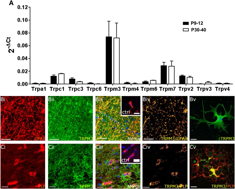
Fig. 4.
Expression of TRP channels in optic nerve glia. a qRT-PCR of acutely isolated optic nerves from WT mice aged P9–P12 and P30–P40; data are from 10 pooled optic nerves in each age group, run in triplicate, expressed as relative mRNA levels (2-ΔCt) compared to the housekeeping gene GAPDH method (mean ± SEM, n = 3). TRPM3 was the most highly expressed TRP channel in the postnatal and adult nerve (***p < 0.001, ANOVA and unpaired t tests) and there was no developmental regulation of TRP channels, which had a rank order of expression in the adult of TRPM3 >>> TRPM7 (***p < 0.001, unpaired t test) > TRPC1 (*p < 0.05, unpaired t test) >> TRPV2 (**p < 0.01, unpaired t test); TRPC3, TRPM6 and TRPV3 were expressed at significantly lower levels, and TRPA1, TRPC6, TRPM4, and TRPV4 were barely detectable. Immunolabelling for TRPM3 (b, c, green) with GFAP to identify astrocytes in WT nerves (b, red) and in PLP-DsRed nerves to identify oligodendrocytes (c, red), in optic nerve sections (bi–iv, ci–iv) and explant cultures (bv, cv). Confocal micrographs illustrate single channels (bi, bii, ci, cii) and merged cannels (biii, ciii). The colocalization channels illustrate voxels in which green and red channels are of equal intensity and appear yellow, showing expression of TRPM3 is localized to astroglial processes (bv) and oligodendroglial somata (cv). Immunostaining of explant cultures shows astrocytes are immunopositive for TRPM3 (bv) and that TRPM3 is localized to oligodendroglial somata and excluded from their distal processes (cv). No immunostaining was observed in negative controls that were pre-incubated in blocking peptides for TRPM3 (insets, biii, ciii). Nuclei are stained with Hoechst blue. Scale bars 20 μm

