
Figure 4.
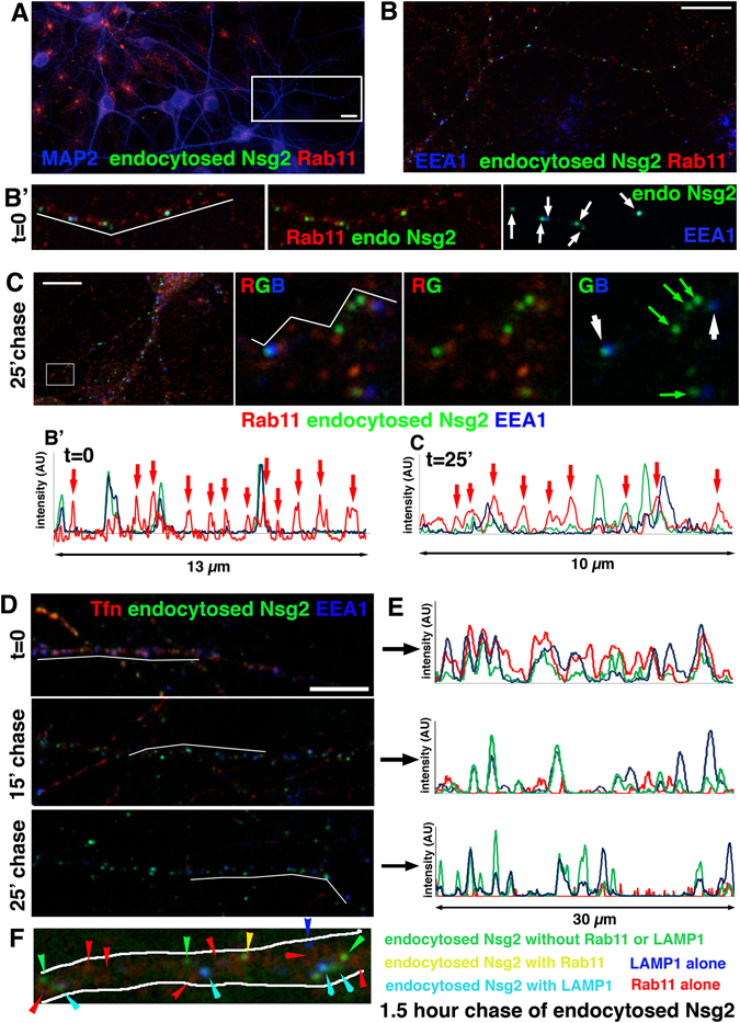
Endocytosed Nsg2 barely enters recycling endosomes. (A–C) An endocytosis assay with endogenous Nsg2 was carried out and cells either fixed (t = 0; A,B) or returned to the incubator for a 25 minute chase period before fixation (C). Bar = 10 µm. After fixation, cells were counterstained for MAP2 to mark dendrites (A), EEA1 to mark early endosomes, and endogenous Rab11 to mark recycling endosomes (B). (B’) shows one dendrite as merged images of all three channels and combinations of two channels as marked on the panels. Endocytosed Nsg2 (green) rarely co-localizes with Rab11 (red; arrowhead), but shows many instances of co-localization with EEA1 (blue; arrows). A line scan along the white line in B’ is shown. (C) After 25 minute chase, endocytosed Nsg2 still shows some co-localization with endosomes containing high levels of EEA1 (white arrows), but no increase in overlap with Rab11 (red) is observed (see red arrows in the corresponding line scan along the white line). Green arrows indicate endosomes containing endocytosed Nsg2 with low levels of EEA1. R = red, G = green, B = blue. Corresponding experiments for endocytosed endogenous Nsg1 are shown in Suppl. Figure S6. (D,E) Cy3-Tfn (red) and anti-Nsg2 antibody (green) were simultaneously fed to live cultures for 30 minutes (endocytosis assay) and then fixed at t = 0, or after 15 minute or 25 minute chase times (D). Bar = 10 µm. After fixation, cells were counterstained with MAP2 to identify dendrites (channel not shown) and EEA1 to identify early endosomes (blue). Since Tfn recycles rapidly, the red channel intensity for the 15 and 25 minute chase time was increased so that localization of the small number of remaining Tfn compartments could be more easily visualized in the panel. (E) Shows the corresponding line scans through the MAP2-positive dendrite shown in (D). Tfn co-localizes extensively with endocytosed Nsg2 and with EEA1 at t = 0, but then rapidly leaves the early endosomes whereas endocytosed Nsg2 remains co-localized with EEA1 throughout the chase. (F) Endocytosed endogenous Nsg2 was chased for 1.5 hours and then counterstained against endogenous Rab11 and LAMP1. A close-up of one representative dendrite is shown. Colored arrowheads correspond to endosomes with presence or absence of the stained markers. The color coding is indicated on the right. Turquoise arrowheads indicate endocytosed Nsg2 present in LAMP1-positive late endosomes/lysosomes.