Fig. 1.
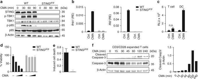
STING activation induces apoptosis in T cells. a CD4+ T cells from wild-type (WT) or STINGgt/gt mice were treated with DMSO or 10-carboxymethyl-9-acridanone (CMA) for the indicated times and phosphorylation of TBK1 and NF-κB (p65) was assessed by immunoblot. b CD4+ T cells from WT or STINGgt/gt mice were treated with DMSO or CMA for 4 h and abundance of mRNAs of the indicated genes was measured by RT-qPCR. c Supernatants from CD4+ T cells or dendritic cells (DC) treated with DMSO or CMA were collected after 16 h. A bioassay was used to measure the production of type I IFNs (RLU relative light units). d, e CD4+ T cells from WT and STINGgt/gt mice were treated with DMSO or CMA. After 16 h cell viability d and percentage of apoptotic cells e, assessed by Annexin V staining, were determined. f WT CD4+ T cells were stimulated with CMA for the indicated times and caspases-3 cleavage was assessed by immunoblot. Quantification of cleaved Caspase-3 relative to β-Actin levels is shown on the right panel. Data are representative of n = 3 (a, f) independent experiments or mean and s.d. of technical replicates (n = 2) of one representative experiment out of n = 3 (b, d, e) or n = 2 (c) independent experiments are shown. Unprocessed original blots are shown in Supplementary Fig. 9.

