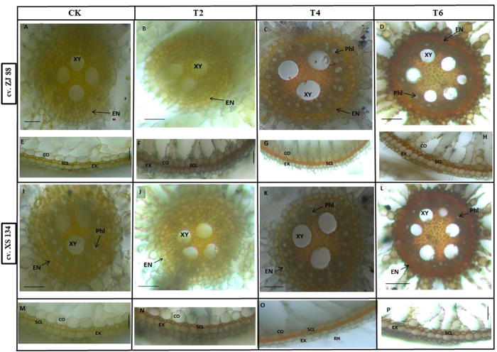
Figure 5.
Comparison of lignin deposition in rice roots. Lignin staining in the central cylinder (CC) (A–D and I to L) and the outer part of the roots (E–H and M–P) grown in salt and 2,4-D treatments. Lignin in the cell walls was detected by an orange/brown staining. Root tips (0.5 cm) were stained with Maule reaction, then cross-sectioned and examined under a light microscopy (LEICA MZ 95 microscope equipped with LECIA DFC 300 FX camera). A slight development of lignin was observed in the central cylinder (CC) of roots under 2,4-D treatments (B,J) in both rice cultivars. Relatively brighter staining in the CC of roots of cultivar ZJ 88 (C,G) compared to XS 134 (K and O) was found under higher saline stress treatment (T4). Under higher saline and 2,4-D combined treatment (T6), staining turned to reddish brown, showing intense lignin deposition in endodermis cell and its adjacent cell layers in cultivar XS 134 followed by ZJ 88. Under 2,4-D alone treatment (F,N) thin, light brown sclerenchyma and exodermal cell walls in outer part of the roots (OPR) of rice cultivars can be found. Under higher saline stress treatment, sclerenchyma cells with faint orange color can be observed in the figures (G,O). Intense reddish brown stains in root of the cultivar XS 134 followed by cultivar ZJ 88 can be seen under combined salt and herbicide stress treatment (H,P). 20-days-old rice seedlings were treated as control (CK) with EC 1.2 dS m−1, T2 (recommended dose of 2,4-D), T4 (EC 8 dS m−1), T6 (EC 8 dS m−1 + recommended dose of 2,4-D) for 15 days. Scale bar = 0.05 mm.

